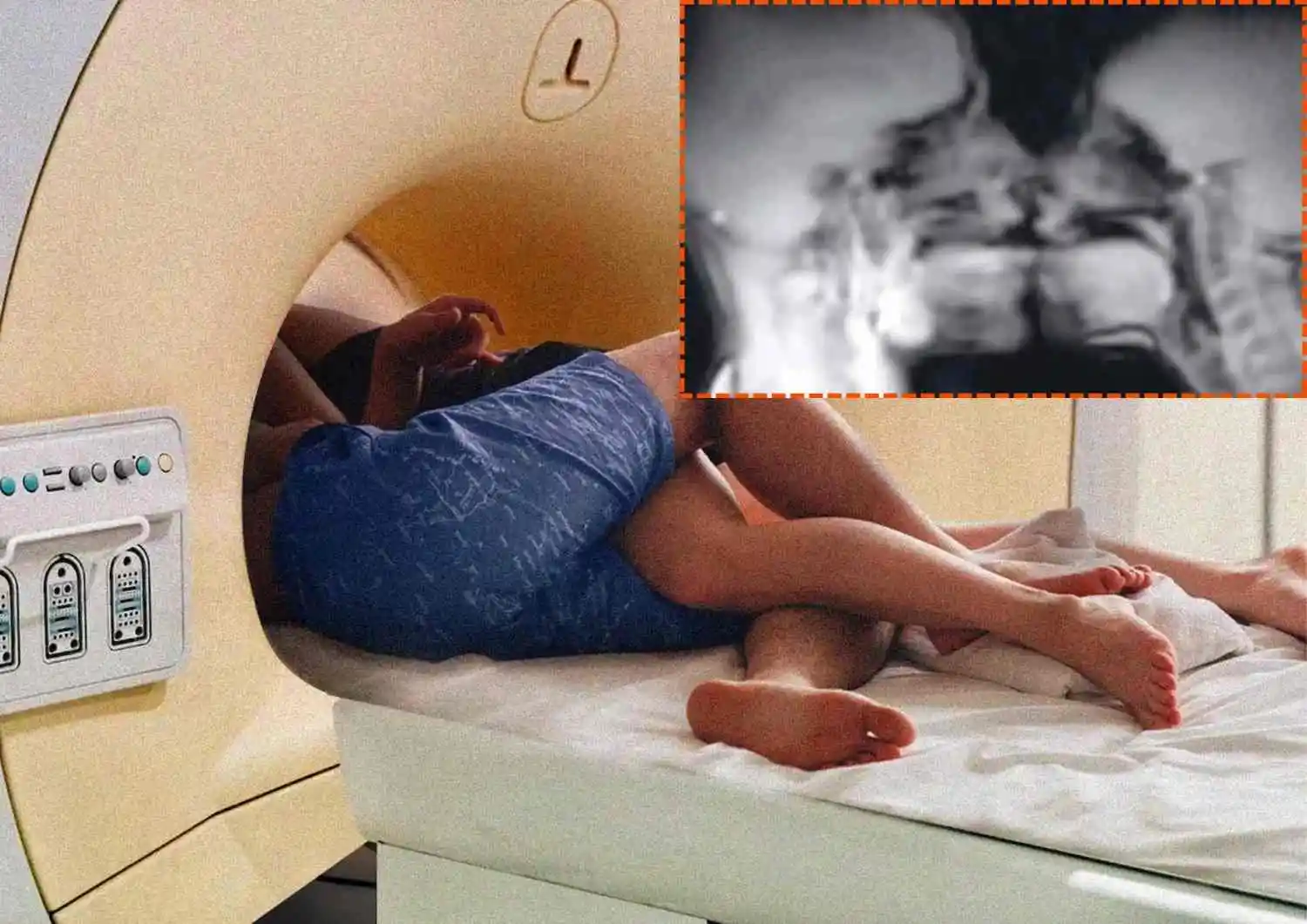
In 1991, researchers at University Hospital Groningen in the Netherlands began an unusual experiment: using magnetic resonance imaging (MRI) to see what happens inside the human body during sexual intercourse. Anthropologist Ida Sabelis and her partner took part in the first successful scans. The research later involved eight couples and three women who were scanned during sexual arousal. The findings were published in the British Medical Journal on December 18, 1999, under the title Magnetic Resonance Imaging of Male and Female Genitals During Coitus and Female Sexual Arousal.
One of the study's most surprising findings concerned the shape of the penis during intercourse in the missionary position. The MRI images showed that it bends into a boomerang shape, with about one-third of its length inside the body. The scans also showed changes in the female body during sexual arousal. The uterus moved upward, while the front wall of the vagina became longer. However, researchers found no increase in the overall size of the uterus.
The study challenged some earlier ideas about human sexual anatomy and showed how MRI technology could help researchers observe the body during activities that are difficult to study. In 2000, the research team received the Ig Nobel Prize in Medicine, an award that recognises research that first makes people laugh and then makes them think. The paper later became one of the BMJ's most downloaded articles, attracting attention for its unusual approach to medical research.